


As a result of a person's upright walking, the spine takes on the main load as an axial structure. Therefore, degenerative and dystrophic processes are quite common consequences of human life. One of the most common diseases of the musculoskeletal system is osteochondrosis, which can cause severe discomfort and disability. This article will discuss the most severe form of this pathology - general osteochondrosis.
general characteristics
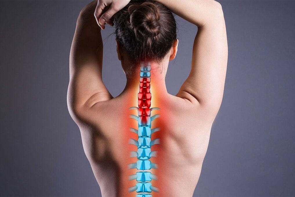
Osteochondrosis is a degenerative disease of the spine, mostly affecting the chest, back and neck regions. This pathology is directly related to age. The disease is more common in people over 40 years of age, but recently there has been a younger trend. General osteochondrosis differs in that it affects more than one department or several departments at the same time. Due to the progressive development of degenerative processes not only in the bone tissue, but also in the ligamentous apparatus of the spine, the vertebrae become mobile and put pressure on the nerves and blood vessels. Symptoms of general osteochondrosis are associated with it, but it is noteworthy that the disease can be asymptomatic for some time.
It is important! Pathology requires multifaceted control, because it affects not only the musculoskeletal system, but also the nervous system, as well as internal organs. In addition to the spine itself, the pathological process can affect other elements of the skeleton.
Etiology and pathogenesis
There are many causes of widespread osteochondrosis. Some of them are associated with congenital skeletal defects, others with inadequate load during vigorous activity. General factors that contribute to the development of the clinical picture in particular:
- injuries;
- straight legs;
- heel foot - deformation of the foot (equinovarus, varus, valgus, depending on the condition of the heel);
- work related to lifting heavy loads;
- playing sports without heating or heating the muscles;
- work at low temperatures.
Low temperatures are considered provoking factors, because cold temporarily changes the molecular structure of soft tissues, reduces the intensity of blood circulation, the permeability of nerve impulses and metabolism, and therefore the activity of the immune system. Other causes disrupt the biomechanics of the spine and contribute to rapid wear of the intervertebral discs.
Pain in widespread osteochondrosis can be the result of osteophytes or disc deformation. The pain is usually radicular, that is. due to compression of the posterior nerve roots.
General osteochondrosis easily mimics other diseases. With damage to the thoracic region, pain appears in the heart region and is mistaken for ischemic processes, and with damage to the lumbar regions, it is mistaken for radiculitis.
Symptoms
Clinical manifestations will depend on which parts are affected and which joint.
When the cervical spine is affected, the following are characteristic:
- unstable blood pressure;
- Headache;
- lack of coordination;
- pain in hands;
- numbness in the upper body and arms.
For pathology of the thoracic region:
- intercostal neuralgia;
- stiffness in arms and neck;
- dysfunction of internal organs.
If the lumbar region is affected:
- burning;
- urinary disorders;
- spasms;
- pain when walking.
Based on the above, it is easy to conclude that the pathology affects not only the spine and large joints, but also the autonomic nervous system. The latter is associated with interruptions in the work of internal organs. General polysegmental osteochondrosis can sometimes worsen. In such cases, the manifestations are much more intense. With a combination of disorders of several departments, the symptoms will be consistent.
Complications
Osteochondrosis can be conventionally divided into moderate osteochondrosis, which is a natural wear process of the spine as a result of life activity, and severe osteochondrosis, which is characterized by the most complications.
Moderate osteochondrosis is easily treated with conservative treatment. And if it is impossible to completely stop the inevitable aging process, it is quite possible to significantly slow it down. Complications that severe osteochondrosis can lead to are as follows.
- Spondyloarthrosis.
- Degeneration of the intervertebral disc.
- Spinal stenosis.
It is important! Intervertebral discs act as shock absorbers and reduce friction between the vertebrae. Degenerative processes in these structures can lead to the protrusion of the nucleus pulposus of the disc and the formation of an intervertebral hernia. Protrusion causes root compression and pain.
Spondyloarthrosis is a degeneration of the facet joints that connect adjacent vertebrae. In another way, such joints are called facet joints. When the articular cartilage is damaged, painful contact occurs between the vertebrae. With the degeneration of the facet joints, most bone growths appear, which causes spondylosis.
Stenosis is a narrowing (in this case of the spinal canal). Typically, stenosis is the result of pathologies such as intervertebral hernia or spondylosis. Bony protrusions and hernial protrusions compress nerve roots at their entry and exit points.
The clinical picture of severe osteochondrosis is the result of complications:
- chronic pain in the spine;
- friction of bone surfaces;
- hardness;
- sudden muscle weakness;
- decreased reflexes;
- tingling in the limbs;
- radiating pain;
- sciatica symptoms.
It is caused by compression of the sciatic nerve.
Classification
There are four degrees of osteochondrosis. Classification is based on the collected history and with the help of instrumental diagnostic methods. The main criteria in this classification are pain and neurological symptoms.
- I degree - the pain is easily relieved by medication.
- Grade II - characterized by long-lasting pain and spinal deformity with moderate neurological symptoms.
- III degree - pain is systematic, neurological symptoms are significant.
- IV degree - constant pain, multiple neurological deficits. Disturbance of the conduction of nerve impulses. Paralysis and paresis.
In the case of widespread dysplastic osteochondrosis, the patient is given the status of disability. Depending on the general condition of the patient, the degree of development and intensity of the clinical picture, disability can be of three groups.
Types of disability in osteochondrosis.
| Group | Description |
|---|---|
| The first group | The functions of the spine are lost. The patient cannot move independently and take care of himself. |
| The second group | The patient is able to move and perform small tasks, but periods of exacerbation are frequent. The operation is contraindicated or useless for some reason. Or he has already been operated on, but it was unsuccessful. |
| The third group | The patient is capable of self-care. There is pain and vestibular symptoms, but the frequency of exacerbations is moderate and periodic. |
The disability group is assigned by a doctor based on some studies to assess the ability to work.
Diagnostics
When visiting a doctor, the diagnosis will consist of several components. The first and most important is to collect anamnesis based on subjective information provided by the patient. Because osteochondrosis has a genetic component, family history is emphasized. The specialist asks about the place of work, living conditions and the disease itself, and the patient must describe exactly what is bothering him. The best results can be achieved with good feedback between the patient and the doctor.
The next method is an objective study carried out by a specialist himself or using instrumental methods. The doctor checks the range of motion of the neck and limbs, which may be noticeably reduced due to pain and stiffness. Using palpation, he notes how spasmed the muscles are and how curved the spine is. Emphasis is placed on the neurological examination, with the help of which the weakened reflexes can be monitored. This symptom may be the result of nerve compression or damage.
Instrumental methods for diagnosing general osteochondrosis include:
- X-ray of the entire spine in two projections.
- MRI to evaluate ligaments and nerve tissue.
- An electrophysiological study to test the conduction of nerve impulses.
X-ray is effective for determining the presence of bone growths - osteophytes, narrowing of the spinal canal and other diseases resulting from osteochondrosis, for example, scoliosis.
Computed tomography can also be used in conjunction with MRI. Using a CT scan, you can determine the degree of nerve compression by spurs.
The diagnosis of widespread polysegmental osteochondrosis is made if other pathologies that cause the destruction of the vertebrae (for example, tuberculosis) are excluded and affect several segments of one or more departments.
There are additional diagnostic methods. These include:
- Bone scan.
- Discography.
- Myelogram.
A bone scan can detect conditions such as osteoarthritis, fractures or infections. This method is a radionuclide and is convenient for differential diagnosis and identification of possible complications.
During discography, a contrast agent is injected into the nucleus pulposus of the intervertebral disc. This method is effective in determining the presence of a herniated disc.
Myelogram is also a contrast research method. Contrast is injected into the spinal canal and the image is recorded using X-ray or CT. Using this method, you can determine the condition of the spinal cord, the presence of constrictions and compressions.
Treatment
Treatment is based on the following mechanisms.
- Slowing down the degenerative process by improving the supply of nutrients to the structures of the musculoskeletal system.
- Stabilization of the spine.
- Elimination of compressed nerve fibers.
- Elimination of symptoms.
The following drugs are used for drug treatment:
- non-steroidal anti-inflammatory drugs that relieve inflammation and pain;
- Anilides relieve pain in the initial stage;
- local analgesics in the form of ointment;
- muscle relaxants to reduce muscle spasm and increase range of motion;
- B vitamins to improve nerve tissue permeability;
- chondroprotectors that reduce the rate of development of degenerative processes by integrating active substances (chondoitin sulfate and glucosamine) into cartilage cells. As a result, metabolism normalizes and clinical manifestations decrease. The drug has been used for a long time, and special consultation is required during pregnancy, lactation, and in the presence of gastrointestinal diseases. An absolute contraindication is phenylketonuria;
- antispasmodics eliminate spasms of smooth muscles and thereby alleviate the manifestations of osteochondrosis in internal organs;
- antioxidants;
- antidepressants to eliminate the psychosomatic component of this disease. They prevent the transmission of nerve impulses from the central nervous system to the brain. Promote the production of endorphins and help solve the problem of chronic insomnia due to constant pain.
- neuropathic agents to eliminate damage to nerve endings.
- opiates for excruciating pain and failure of other pain relievers.
The following are used as invasive medical procedures:
- injection of steroids into the epidural space. Steroids are powerful anti-inflammatory drugs. They relieve inflammation of the nerve roots, which helps relieve pain caused by radiculopathy. Complex procedure. A qualified specialist is required;
- facet joint injections. Injected drugs cause local numbness and pain relief.
Important to know! Taking medication is not intended to get rid of the disease - there are no drugs that can completely eliminate osteochondrosis, which is a chronic disease. Medicines are prescribed only to relieve symptoms.
Medicines are prescribed by the attending physician. The patient is informed about the possible side effects of each drug and then decides for himself which course of treatment to choose.
For symptoms that cause suspicion of osteochondrosis, contact a vertebrologist, orthopedist and neurologist. High-quality medical care will consist of close cooperation of these specialists with each other and with the patient.
Physiotherapy
Physiotherapy is used as a complex of auxiliary therapeutic measures to improve blood circulation and metabolism in the affected tissue. The following methods are used for widespread osteochondrosis.
- Electrophoresis (based on the movement of colloidal particles under the influence of an external electric field).
- Phonophoresis (combination of ultrasound and drugs).
- Magnetotherapy (use of static magnetic field).
- UHF therapy (ultra high frequency therapy).
- Electromyostimulation (nerve and muscle stimulation).
- Acupuncture (acupuncture).
- Laser exposure.
In addition to physiotherapy, manual therapy and physical therapy are actively used. Professional massage can lead to long-term remission. During the exacerbation of the disease, therapeutic exercises should not be performed, as this can lead to complications. During the period of remission, moderate physical activity maintains muscle tone and, therefore, the spine. Exercises are conducted under the supervision of an instructor and prescribed by the attending physician.
During exacerbation, you can not warm your waist, but you can wear a corset, but only for a few hours. In other cases, wearing a corset for more than a few hours is not optimal, as it can cause muscle atrophy.
Manual therapy can increase clearance of pinched nerves and reduce neurological symptoms. Alternative methods are leeches and vacuum massage. These methods are aimed at improving blood circulation in the affected area. Sanitation-spa treatment is useful. Special preference is given to water procedures.
Surgery
When treating osteochondrosis, specialists are more willing to resort to conservative therapy, but for the correct effect, a lot of time, patience and strict adherence to the patient's recommendations are necessary. If conservative treatment is ineffective, only then should you resort to invasive methods. Usually the operation is palliative. This means that the operation will be performed only to relieve symptoms and relatively improve the quality of life, but not for complete recovery (in fairness, it should be noted that conservative treatment does not lead to complete elimination of the disease, but to the acceptance of the patient. It is better to switch to non-invasive therapyis a sign of prognosis).
There are two types of operation: decompression and stabilization. The first is aimed at relieving nerve compression, and the second is aimed at stabilizing the spine. The following operations are classified as decompression operations.
- Facetectomy - removal of the facet joints to relieve compression.
- Foraminotomy is an increase in the lumen of the narrowed spinal canal due to osteophytes.
- Laminectomy is the removal of the back part of the vertebra, which may be deformed due to osteochondrosis.
- Laminotomy - removal of a piece of the back of a vertebra to widen the spinal canal.
These operations require a posterior approach, but in the case of an intervertebral hernia, the surgical approach will be anterior.
Decompression operations with an anterior approach are as follows.
- Discectomy - removal of the intervertebral disc.
- Corpectomy - removal of the entire vertebral body with adjacent discs.
Stabilization operations include:
- Spinal fusion is a method of joining the vertebrae together.
- Artificial intervertebral disc.
The need for stabilizing operations arises after discectomy.
Surgical interventions are rarely prescribed, because there is a risk of developing serious complications.
Complications include:
- recurrence of pain;
- false union;
- infection;
- phlebitis in limbs;
- urinary disorders;
- pain due to the graft;
- failure of installed fasteners.
The postoperative period lasts several months. Stitches heal 2 weeks after surgery. If complications are detected, you should immediately consult a doctor.
After the operation, a rehabilitation course is conducted to speed up the healing process and restore full working capacity.
General recommendations
Proper nutrition helps prevent relapses of exacerbations of general osteochondrosis. Proper nutrition is primarily necessary to maintain a stable body weight, as excess weight puts additional stress on the spine. In this case, the diet should be complete, enriched and rich in calcium, magnesium and potassium. Coffee consumption should be limited as it leaches calcium from the body. It is useful to visit the pool. You should always avoid staying in one position.
If there are frequent exacerbations and a lack of discipline to strictly follow the recommendations, it is better to undergo a full course of treatment in a hospital under the supervision of a doctor.
You cannot take medicine yourself.
Let's summarize
Often, widespread osteochondrosis develops after incompletely treated "single" osteochondrosis. This fact shows that if any discomfort arises, you should immediately consult a doctor without hoping that the pain will go away by itself. In this case, it will be easier to prevent the development of other pathologies and to treat the root cause.